Introduction
Useful data regarding the safety and efficacy of perispinal etanercept (PSE) for chronic stroke may be developed through properly designed randomized clinical trials (RCTs). Poorly designed clinical trials, however, may yield incomplete, misleading, or inaccurate data that do not reflect the clinical results observed in clinical practice. The design of clinical trials for innovative treatments, such as perispinal etanercept for chronic stroke, may be particularly difficult, particularly for physicians with limited experience utilizing perispinal etanercept for chronic stroke in the clinic. Fortunately now, in September 2025, the results of two randomized clinical trials studying the safety and efficacy of perispinal etanercept for chronic stroke have been performed. The results of the first double-blind, placebo-controlled randomized trial testing the safety and efficacy of perispinal etanercept for chronic stroke was completed in 2019 and published in January 2020. In this first trial remarkable and unprecedented reduction in intractable central post-stroke pain and significant improvement in shoulder mobility was documented in the participants who received perispinal etanercept, and both improvements were statistically significant in comparison with placebo. This first trial was conducted using the basic trial design conceived by Dr. Edward Tobinick, the inventor of the perispinal etanercept for chronic stroke treatment.
The published results of the second trial designed to provide data regarding the safety and efficacy of perispinal etanercept for chronic stroke were first made available on September 4, 2025. These results were from the Perispinal Etanercept to Improve Stroke Outcomes (PESTO) clinical trial. This trial did not use a design by Dr. Tobinick. The trial design of PESTO has been severely criticised. To understand why, one needs to start with an understanding of the scientific background. What follows is a review of basic background material, followed by a detailed discussion of the optimal design for clinical trials testing perispinal etanercept for chronic stroke.
TNF (tumor necrosis factor) is an immune signaling molecule that plays a pivotal role in the modulation of synaptic mechanisms in the brain[1-5]. Stroke results in chronic microglial activation and elevation of TNF in the brain, a disturbance of homeostasis that may self-perpetuate through an autocrine feed-forward loop[6-12]. Clinical and imaging evidence suggests that microglial activation is widespread in the brain and persists long after the acute stroke[10-16]. The 18-kDA translocator protein (TSPO), previously named the peripheral benzodiazepine receptor (PBR), is a mitochondrial protein that is markedly increased at sites of brain injury and is considered a marker of microglial activation[17,18]. Radioligands targeting TSPO have been used with positron emission tomography (TSPO PET) to image microglial activation in the brain after stroke, in humans and in basic science models[10,11,15,17,18].
Perispinal etanercept is an innovative biological treatment designed to improve recovery from stroke by neutralizing excess TNF and reducing microglial activation[13]. Perispinal etanercept has been used to improve neurological recovery after stroke since 2010[13,14,19]. Real-world evidence, derived from favorable clinical experience involving thousands of patients with chronic stroke treated with perispinal etanercept over the course of 14 years; basic science evidence; expert opinion; imaging studies and a double-blind, placebo-controlled RCT published in 2020 all support the use of perispinal etanercept to reduce chronic post-stroke neurological dysfunction and improve recovery from stroke[8,12-16,19-28]. However, formidable barriers to widespread acceptance exist, particularly for novel treatments that challenge existing dogma[27,29-35]. It is well known that an incorrect design of an early phase RCT could result in the failure of an effective treatment to reach those who could benefit[36-40]. Such a false negative result would be considered a Type II error, i.e. the failure to detect the true efficacy of an effective drug.
A primary consideration in the design of chronic stroke RCTs is the fact that the population of individuals with chronic post-stroke neurological dysfunction is inherently heterogeneous[39]. Neurological outcome and the magnitude of response to treatment in chronic stroke is influenced not only by the size and location of the lesion, but also by the type and subtype of stroke – ischemic (thrombotic, embolic, lacunar) or hemorrhagic (intraparenchymal hemorrhage, intraventricular hemorrhage, subarachnoid hemorrhage, hemorrhagic transformation, etc.), as well as by external factors, such as the duration and severity of hypoxia, hypotension or hypoperfusion. This heterogeneity can present a particular challenge for small or even medium-size RCTs in which heterogeneity of the RCT study population could reduce the trial’s ability to detect a beneficial treatment effect. Generic outcome measures meant to quantitate response to treatment for heterogeneous, non-stroke populations, such as the SF-36 quality-of-life measure, may have significant issues regarding validity when used as primary outcome measures for stroke trials[41]. Fortunately there are available strategies, including the use of a prospectively enriched study population, which can improve the validity and the reliability of the efficacy conclusions of chronic stroke RCTs[39,42]. Trial enrichment is defined as “[t]he prospective use of any patient characteristic to select a study population in which detection of a drug effect (if one in fact is present) is more likely than it would be in an unselected population[42].”
To add historical perspective, both infliximab and etanercept failed in early indications, such as sepsis, when they were first studied, more than 20 years ago[43,44]. Both drugs were repurposed and successfully used for the new indications for which they are now FDA-approved, including rheumatoid arthritis, psoriasis, and inflammatory bowel disease. From these early failures it appears that, at least for some of these disorders, such as sepsis, the time point within the natural history of the disease when the TNF antagonist is administered may be critical. For sepsis, the inflammatory cascade initiated by gram-negative bacteremia may be so rapid that by the time the patient is in septic shock it is too late to intervene with a TNF antagonist. For chronic stroke, clinical experience indicates that time to intervention is not critical, since patients respond favorably weeks, months, years or even decades after stroke or brain injury[13,14,19,22,45]. Part of the difference is that in the brain TNF plays a pivotal role in the modulation of synaptic function[1-5,25,46,47]. TNF’s key role in the regulation of brain function is distinct from its role as the master regulator and initiator of peripheral inflammatory cascades, which if unchecked, may eventuate in a cytokine storm[5,9,24,25,27,47-62]. This distinction is certainly part of the explanation why the required timing of TNF inhibition for these different categories of diseases is not identical.
The perispinal etanercept chronic stroke RCT guidelines detailed herein are formulated based upon clinical experience using perispinal etanercept for more than 5,000 chronic stroke patients. Although this review will focus on perispinal etanercept RCT design, the basic concepts may assist those designing clinical trials for other neurological indications.
PSE for Stroke Recovery: Scientific Background
The scientific background supporting the clinical use of perispinal etanercept for brain disorders has been reviewed in detail in previous publications[13,14,21-25,27,45-47,51,55,59,63-72]. The following is a current synopsis of selected aspects of the scientific background.
TNF
TNF is an immune signaling molecule and a neuromodulator that plays essential roles in immune and inflammatory responses throughout the body[55,73]. In the periphery, TNF is the master regulator of the inflammatory response[24]. In the brain, TNF is a neuromodulator that regulates synaptic and brain network function, including synaptic transmission, synaptic strength, synaptic scaling and synaptic plasticity[1-3,5,21]. Elevated brain levels of TNF are thought to be centrally involved in the pathogenesis of the chronic brain dysfunction that accompanies stroke, traumatic brain injury, Alzheimer’s disease and other brain disorders by mediating synaptic dysfunction and perpetuating microglial activation[9,13-15,19,21,22,24,25,27,45-47,55,69,74-77].
Etanercept
Etanercept is a dimeric fully human soluble TNF receptor Fc fusion protein manufactured using recombinant DNA biotechnology. It is a large molecule composed of 934 amino acids with a molecular weight (mw) of 150 kilodaltons (kDa). It consists of the extracellular ligand-binding portion of the human 75 kilodalton (p75) TNF receptor linked to the Fc portion of human IgG1. Etanercept reversibly binds to circulating TNF, acting as a decoy receptor, reducing the biologic effects of TNF, including TNF’s deleterious effects when present at levels in excess of its normal physiologic range. Etanercept’s first Food and Drug Administration (FDA) approval was in November 1998 for treatment of rheumatoid arthritis. Etanercept was subsequently approved for chronic use by once or twice weekly subcutaneous injections for multiple additional indications, including ankylosing spondylitis, psoriasis, psoriatic arthritis and juvenile arthritis for children as young as age 2. It has also been used off-label for multiple inflammatory conditions, including several neurological disorders. Since its introduction more than two decades ago it has maintained a favorable safety profile[78,79].
Stroke may result in widespread, sustained microglial activation in the brain
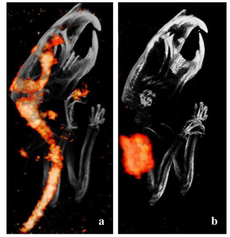
A growing body of evidence suggests that post-stroke microglial activation may persist in the stroke penumbra and in regions remote from the region of acute necrosis after both ischemic and hemorrhagic stroke[8,12,16,80,81]. This evidence includes the results of a 2021 positron emission tomographic imaging study (TSPO PET) of individuals with chronic middle cerebral artery (MCA) stroke that was conducted using a radioligand to the 18 kDa translocator protein (TSPO)[12]. TSPO is a mitochrondrial protein upregulated in activated glia and considered a marker of neuroinflammation[12]. The study found evidence of glial activation in multiple non-infarcted brain regions, both ipsilateral and contralateral, outside the MCA infarct zone, consistent with previous TSPO PET clinical imaging studies[10-12].
Etanercept reduces neuroinflammation by reducing microglial activation and neutralizing excess TNF
Autocrine activation of microglia by TNF creates a positive feed-forward loop that may perpetuate neuroinflammation and have neurotoxic consequences[7,9,13,82,83]. Etanercept reduces neuroinflammation in two ways:
-
By attenuation of microglial activation through a direct effect on microglia and by interruption of the known microglia/TNF autocrine feed-forward loop[7,9,13,57,60,84-96]; and
-
By binding to soluble TNF and thereby reducing its biological activity [97,98].
In addition, neutralization of TNF by etanercept would be expected to reduce the concentration of other non-TNF inflammatory cytokines that could activate microglia because TNF sits on top of the inflammatory cascade as the master regulator of the inflammatory response[9,52-55,99-101]. The inflammatory cytokines IL-1, IL-6, etc. are downstream in the inflammatory cascade from TNF[9,52-55,99-101].
Known physiological mechanisms are consistent with the rapid and sustained neurological improvement
Etanercept has been found to reduce microglial activation in at least 17 animal models, including those studying stroke, as previously reviewed[14,57,60,72,84-96]. Interruption of the autocrine microglia-TNF feed-forward loop may explain the sustained and sometimes progressive neurological improvement that has been observed in chronic stroke patients after a single dose of perispinal etanercept [13,14,19]. The nearly instantaneous attenuation of the biological activity of TNF in the presence of etanercept, and TNF’s known pivotal role in the modulation of synaptic mechanisms, provide mechanisms to explain the rapid neurological improvements seen in stroke and other neuroinflammatory conditions following perispinal etanercept [1-5,14,19,21,22,45,72,75-77].
The Cerebrospinal Venous System
The veins, venous sinuses and venous plexuses of the brain and the spine together comprise the cerebrospinal venous system (CSVS), a unique, large capacity, interconnected, venous system that carries venous blood back and forth between its cerebral and spinal components[102-105]. The first of the two main divisions of this system, the intracranial veins, includes the cortical veins, the dural sinuses, the cavernous sinuses, and the ophthalmic veins. The second main division, the vertebral venous system, includes the vertebral venous plexuses, which course longitudinally up and down the entire length of the spine. The intracranial veins richly anastomose with the internal vertebral venous plexus (IVVP) in the suboccipital region. The external vertebral venous plexus (EVVP), the only division of the CSVS that contains valves, drains the anatomic region posterior to the spine and directs blood flow internally into the IVVP[106]. Venous blood flow within the IVVP can be bidirectional, due to the IVVP’s lack of venous valves[102-105]. Drugs or contrast delivered into areas that drain into the EVVP may flow cranially into the cerebral venous system via the IVVP[103-105]. The CSVS plays an important role in the regulation of intracranial pressure by facilitating venous flow into and out of the brain with changes in posture. The CSVS provides a direct vascular route enabling the rapid delivery of etanercept to the brain after perispinal injection[13,14,19-22,45-47,51,69,72,75,76,102,105,107].
Perispinal injection enables rapid delivery of drugs to the brain via the CSVS
The scientific rationale underlying the perispinal delivery method is supported by basic science experiments[20,26]; clinical investigations[21,105,108]; clinical trials[28,51]; and cadaver studies dating back more than 200 years, with further anatomic evidence developed in the 1940’s and in the last decade[102-105,109-112]; as well as 25 years of clinical experience[13,14,19,22,28,45,51,63-66,72,75,76,105]. The proper performance and implementation of this technique requires expertise and hands-on training with an expert[47,113].